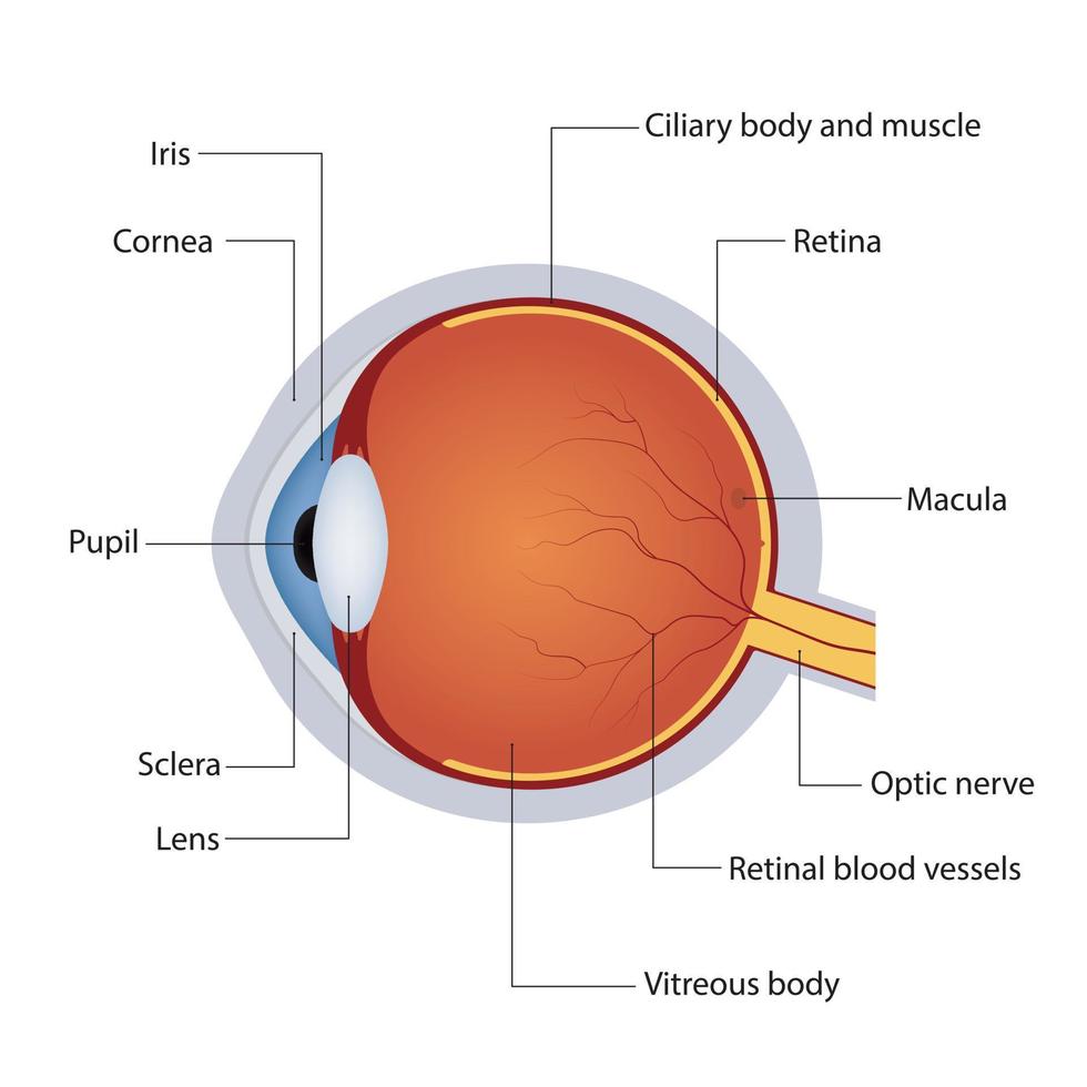
Anatomy of the human eye Colour Theory Understanding and Working Biology Diagrams Learn about the eye anatomy diagram, a spherical organ that detects light and transmits visual information to the brain. The diagram shows the layers, chambers, components, and accessory structures of the eye, as well as its location and function. Learn about the anatomy and functions of the eye, as well as common eye conditions and treatments. See diagrams, photos, and descriptions of the iris, cornea, pupil, lens, retina, and more. Learn about the different parts of the eye, how they work together to see, and what can go wrong. Explore the cornea, iris, pupil, lens, retina, and more with diagrams and explanations.
Learn eye anatomy and functions with interactive figures for medical students. Explore diagrams of the whole eye, external landmarks, cross-sections, retina, brainstem, eyelids, pathways, nerves, muscles and more. Learn about the different parts of the eye and their functions in vision. See diagrams and descriptions of the conjunctiva, sclera, iris, pupil, cornea, uvea, retina, eye muscles, and macula lutea. Learn how the human eye works like a digital camera and see an illustration of its main structures. Click on the eye diagram to get more details about each part of the eye and its function.

Ultimate Guide to Human Eye Anatomy: Parts, Names & Diagrams Biology Diagrams
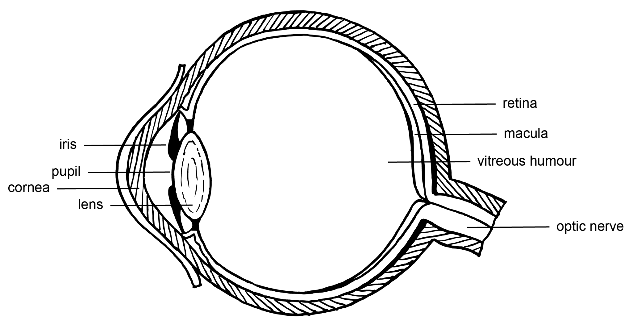
In this interactive, you can label parts of the human eye. Use your mouse or finger to hover over a box to highlight the part to be named. Drag and drop the text labels onto the boxes next to the eye diagram. If you want to redo an answer, click on the box and the answer will go back to the top so you can move it to another box. Learn about the main parts of the eye and how they work together to produce vision. See a diagram of the eye and get tips on eye care services and solutions.