
Brainstem Anatomy Diagram Nervous system Brain angle text png Biology Diagrams Anatomy. The brain stem is a tube-shaped mass of nervous tissue a little over 3 inches (8 cm) long. It is located at the base of the brain, superior to the spinal cord and inferior to the cerebrum. As the brain stem ascends from the spinal cord, it widens and becomes more complex in its structures, both internally and externally. Get a diagram of human brain anatomy and key facts about this important organ. Home . Science Notes Posts; cerebellum, and brainstem, but these portions contain many key sections. and interactions with the world around us. Here is a look at the intricate anatomy of the brain, its functions, and the consequences of damage to different areas. The brainstem is the structure that connects the cerebrum of the brain to the spinal cord and cerebellum. It is composed of three sections in descending order: the midbrain, pons, and medulla oblongata. It is responsible for many vital functions of life, such as breathing, consciousness, blood pressure, heart rate, and sleep. The brainstem contains many critical collections of white and grey

The brainstem is a vital structure that connects the brain to the spinal cord and controls many essential life-sustaining functions. It consists of three main parts: the midbrain, pons, and medulla oblongata. The brainstem acts as a conduit for motor and sensory pathways between the brain and body and contains nuclei that control basic autonomic processes such as heart rate, breathing, and

Neuroanatomy, Brainstem Biology Diagrams
Brainstem death means your brainstem stops functioning. It occurs when something permanently damages the brainstem or cuts off your brain's blood or oxygen supply. Because your brainstem controls essential life functions, you won't be able to regain consciousness. You'll need artificial life support to remain alive.

• Recognize the major internal and external landmarks on the dorsal and ventral surface of the brain stem, so that you can determine if a gross or stained cross section is medulla, pons or midbrain. • Identify on a typical cross section all the brain stem nuclei containing motor neurons that end on striated muscle.
Brainstem: What It Is, Function, Anatomy & Location Biology Diagrams
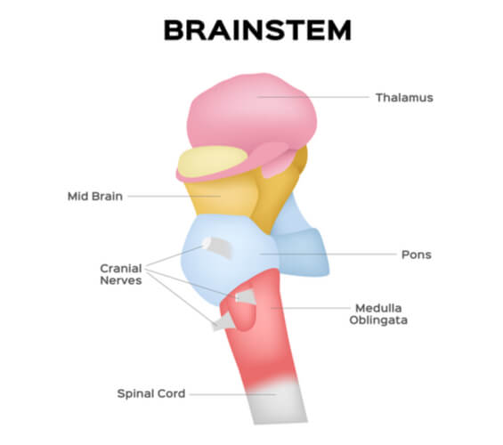
The brainstem is a critical, stalk-like structure situated at the base of the cerebral hemispheres. It serves as the primary conduit for communication between the cerebrum and the rest of the body. Connecting the cerebrum to the spinal cord and cerebellum, the brainstem regulates essential functions such as breathing, consciousness, blood pressure, heart rate, and sleep. Think of the brainstem Brainstem Anatomy: Structures of the brainstem are depicted on these diagrams, including the midbrain, pons, medulla, basilar artery, and vertebral arteries. The medulla oblongata (myelencephalon) is the lower half of the brainstem continuous with the spinal cord. The brainstem (brain stem) is the distal part of the brain that is made up of the midbrain, pons, and medulla oblongata.Each of the three components has its own unique structure and function. Together, they help to regulate breathing, heart rate, blood pressure, and several other important functions.All of these brainstem functions are enabled because of its unique anatomy; since the brainstem
