
Cross Section Anatomy Biology Diagrams Anatomy Atlases is a digital library of human anatomy information. Atlas of Human Anatomy in Cross Section Illustrated Encyclopedia of Human Anatomic Variation Atlas of Microscopic Anatomy - A Functional Approach Anatomy of First Aid - A Case Study Approach Lessons From a Bone Box. Last revised on January 1, 2025 Related Digital Libraries History of Sectional Anatomy. Although it cannot be stated with assurance who first began the study of sectional anatomy, it is certain that the method was in use in the early sixteenth century. Examples of anatomical sections can be found in the anatomical drawings of the Italian genius, anatomist, and artist Leonardo da Vinci (Figs. 1-5).
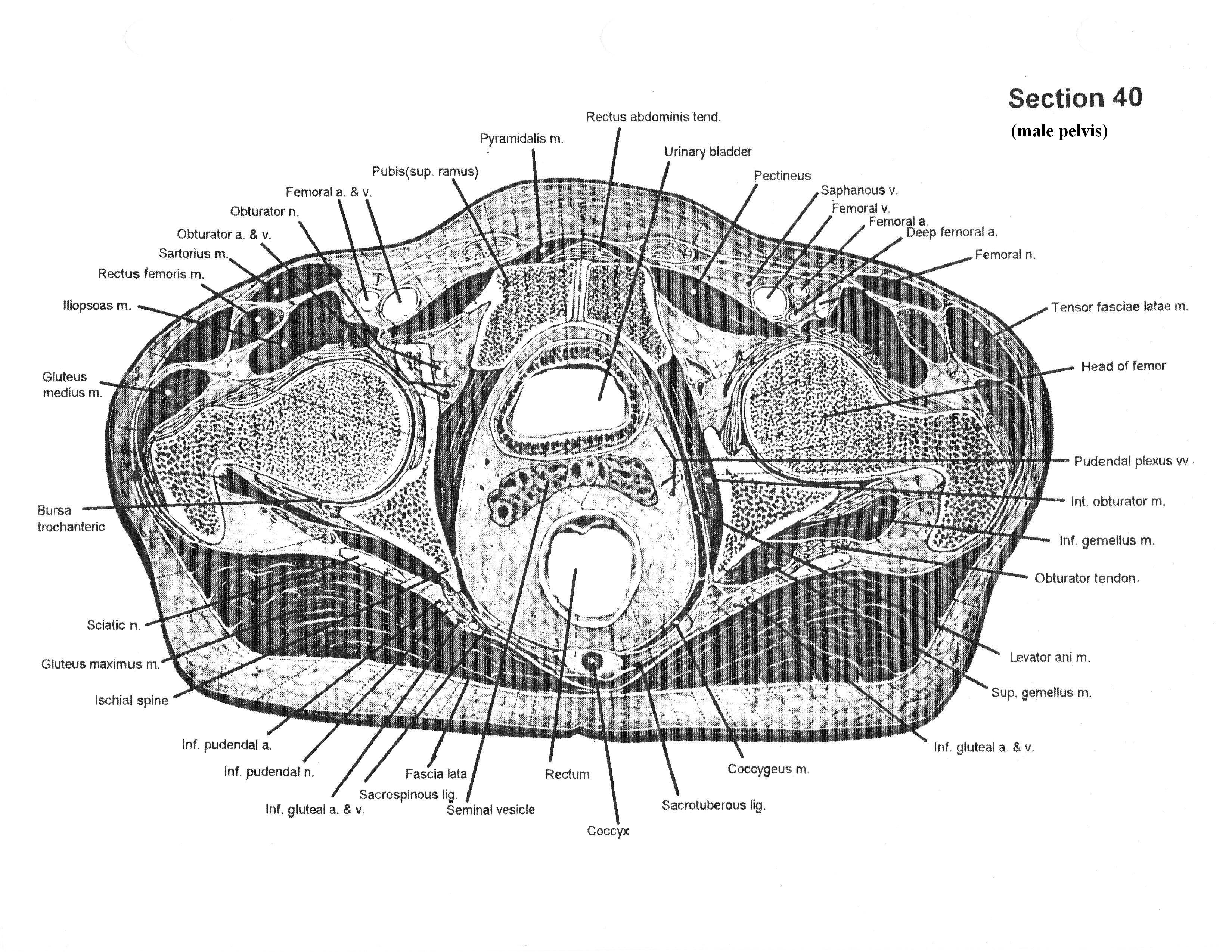
This module presents the anatomy of the whole human body based on cross-sectional photographs of a male cadaver. 1300 anatomical structures have been labeled on 463 photographs of axial cross-sections. This atlas is based on the Visible Human Project ran by the U.S. National Library of Medicine (NLM) under the direction of Michael J. Ackerman. Cross Sectional Anatomy, human anatomy, anatomical sections, CT scan, computed axial tomography, MRI scan, Use scroll bar to go to section of interest You may enlarge any image by 'clicking' on it. Other Websites by Tim Dutra: TransfusionOrders.com & ÓrdenesParaTransfusiones.com Upcoming Websites:

Visible Human Project: Anatomy of the whole human body Biology Diagrams
An atlas of cross sectional human anatomy. Atlas of Human Anatomy in Cross Section: Ronald A. Bergman, PhD Professor of Anatomy Department of Anatomy. Adel K. Afifi, M.D. Section 1. Head and Neck (Plates 1.1 to 1.26) Section 2. Neck, Shoulders, Upper Arm, and Upper Thorax (Lungs) (Plates 2.1 to 2.10)
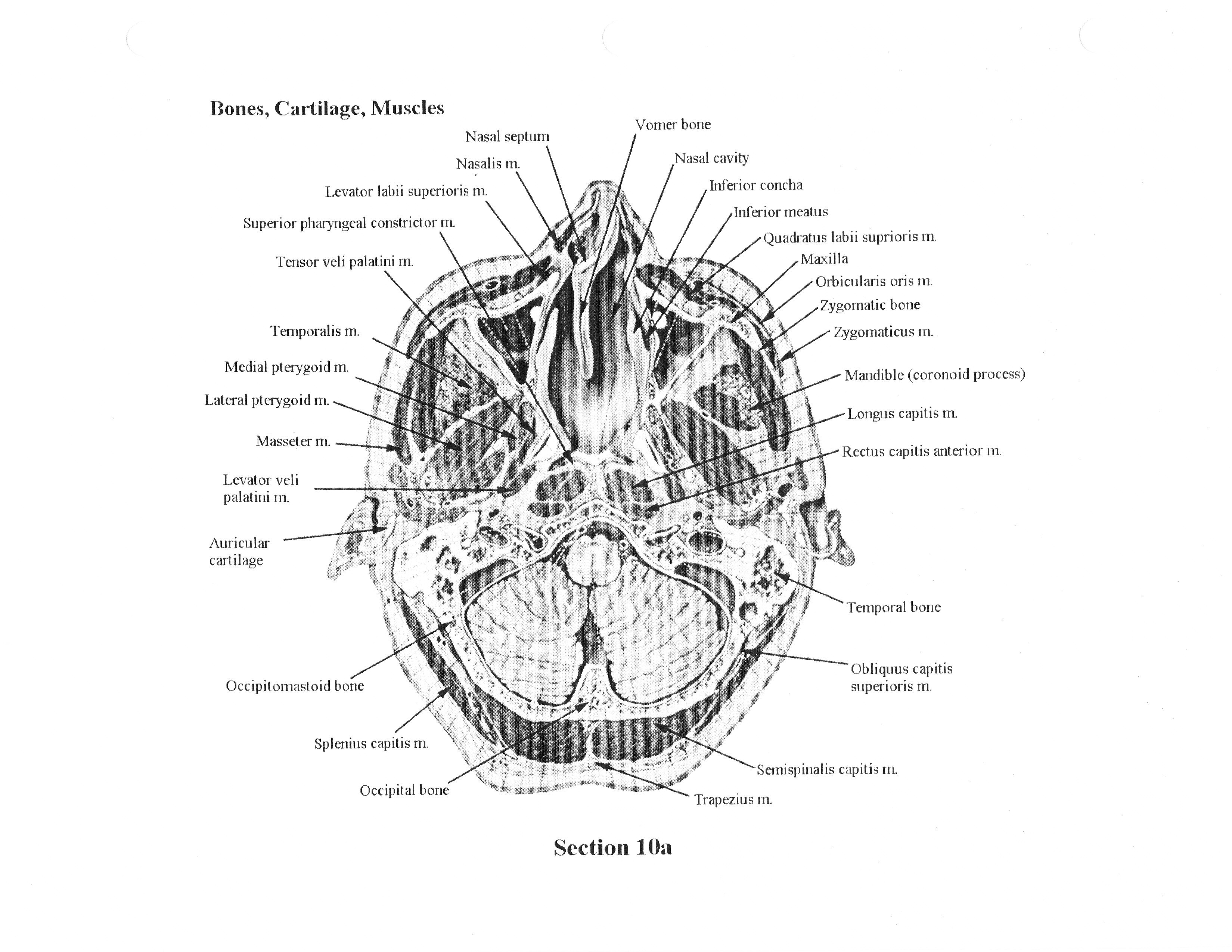
The approximately 7.5 megabyte axial anatomical images are 2048 pixels by 1216 pixels, with each pixel being .33mm in size, and defined by 24 bits of color. The anatomical cross-sections are at 1mm intervals to coincide with the CT images. There are 1,871 cross-sections for both CT and anatomical images.

Anatomy Atlases: A digital library of human anatomy information ... Biology Diagrams
Pectoralis minor muscle (cross-section view) While drawings and paintings using perspective might appear sharp and clearly defined, anatomy is usually quite the opposite. The challenge lies in the fact that the human body is such an intricate machine that anatomical structures, more often than not, are not clearly delimited.

Also known as a cross-sectional plane. Ventral/Dorsal-Equivalent to belly-side and back-side of a body in anatomical position. For a human in anatomical position, this pair of terms is equivalent to anterior and posterior. However, for four-legged animals in what is considered their anatomical position, the belly-side is not equivalent to

Definition and importance of cross sectional anatomy. Biology Diagrams
The Visible Human Project has generated over 18000 digitized sections of the human body. This introduction and tour uses images and animals from the project to teach key concepts in human anatomy. Cross-sectional Anatomy: Using 2D images to visualize 3D structures. Planes of Section, with animations: An introduction to the three planes of Orientation of cross sections Before diving into the deep end, it's important to understand the general orientation of axial anatomy. Every single cross section is viewed from the feet of the patient in a supine position (lying horizontally on his/her back).This means that structures on the right side of the patient's body will be on the left side of the cross-sectional image, and vice-versa.
