
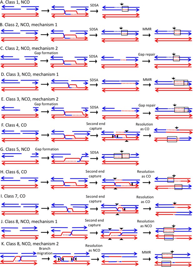
Schematic representation of the recombination event observed in the Biology Diagrams A number of systems have been devised to detect or select for mitotic recombination. In this issue of PLoS Genetics, Lee et al. describe a novel system that represents a major step forward in the study of spontaneous mitotic recombination events. Their studies have given us new insights into the why, when, how, and where of mitotic recombination. Second, a substantial number of mitotic recombination events are associated with a G1-induced DSB, which results in two broken chromatids. The repair of one of the breaks using the homolog as a template might facilitate the repair of the second break by a genetically-silent sister chromatid event, which would inflate the crossover frequency. Mitotic cells favor recombination events that lead to noncrossover events likely to avoid potentially detrimental consequences of loss of heterozygosity and translocations. Figure 1. Open in a new tab. Primary pathways for homology-dependent double-strand break (DSB) repair. Recombinational repair of a DSB is initiated by 5′ to 3′ resection

Mitotic recombination differs from meiotic recombination in several aspects. Recombination is pre-programmed and essential during meiosis, but mitotic recombination operates as a repair mechanism for sporadic DNA damage. Mitotic recombination is an infrequent event (in the order of 10 −7 to 10 −4 per locus per generation). In contrast Second, a substantial number of mitotic recombination events are associated with a G1-induced DSB, which results in two broken chromatids. The repair of one of the breaks using the homolog as a template might facilitate the repair of the second break by a genetically silent sister chromatid event, which would inflate the crossover frequency.
Mitotic Recombination: Why? When? How? Where? Biology Diagrams
Mitotic recombination is a genetic technique used in neuroscience to analyze cell lineages, study gene functions, and identify gene functions. single mitotic recombination events, but also reciprocal products of a single event in the recombinant progeny of males [13]. Once transmitted to progeny, the event becomes fixed, allowing for
Mitotic cells favor recombination events that lead to noncrossover events likely to avoid potentially detrimental consequences of loss of heterozygosity and translocations. Open in a separate window. Figure 1. Primary pathways for homology-dependent double-strand break (DSB) repair. Recombinational repair of a DSB is initiated by 5′ to 3 Mitotic recombination can happen at any locus but is observable in individuals that are heterozygous at a given locus. If a crossover event between non-sister chromatids affects that locus, then both homologous chromosomes will have one chromatid containing each genotype. The resulting phenotype of the daughter cells depends on how the chromosomes line up on the metaphase plate.
Cell Biology of Mitotic Recombination Biology Diagrams
A number of systems have been devised to detect or select for mitotic recombination. In this issue of PLoS Genetics, Lee et al. describe a novel system that represents a major step forward in the study of spontaneous mitotic recombination events. Their studies have given us new insights into the why, when, how, and where of mitotic recombination.

These physiological mitotic death events are promoted in part through a poorly understood and non-canonical mechanism we term MAD-telomere deprotection 22,24,25,26.
